Welcome to the Amira-Avizo Software Use Case Gallery
Below you will find a collection of use cases of our 3D data visualization and analysis software. These use cases include scientific publications, articles, papers, posters, presentations or even videos that show how Amira-Avizo Software is used to address various scientific and industrial research topics.
Use the Domain selector to filter by main application area, and use the Search box to enter keywords related to specific topics you are interested in.
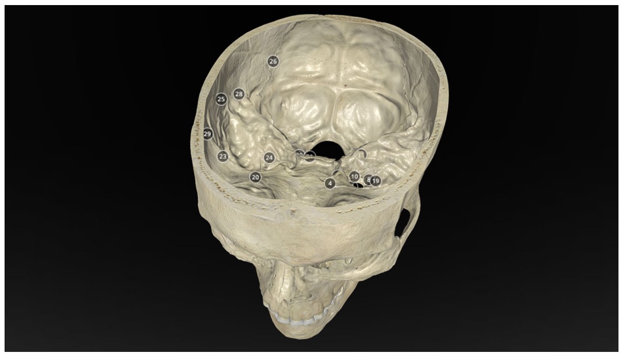
Operative Anatomy of the Human Skull: A Virtual Reality Expedition
Benjamin K Hendricks, MD Akash J Patel, MD Jerome Hartman Mark F Seifert, PhD Aaron Cohen-Gadol, MD, MSc, MBA
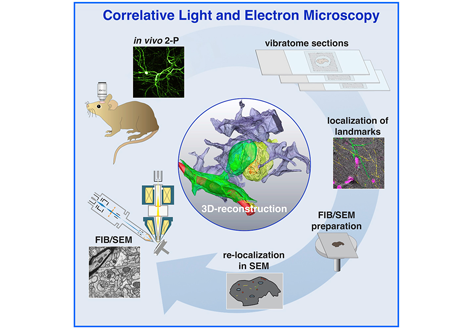
Label-free 3D-CLEM using endogenous tissue landmarks
We demonstrate feasibility of the workflow by combining in vivo 2-photon microscopy and focused ion beam scanning electron microscopy (FIB/SEM) to dissect the role of astrocytic coverage in the persistence of dendritic spines.
Emerging 3D correlative light and electron microscopy (CLEM) approaches enable studying neuronal structure-function relations at unprecedented depth and precision. However, established protocols for the correlation of light and electron micrographs rely ... Read more
Manja Luckner,Steffen Burgold, Severin Filser, Maximilian Scheungrab, Yilmaz Niyaz, Eric Hummel, Gerhard Wanner, Jochen Herms

Cell-type specific innervation of cortical pyramidal cells at their apical tufts
We investigated the synaptic innervation of apical tufts of cortical pyramidal cells in a region between layers 1 and 2 using 3-D electron microscopy (3D-EM) applied to four cortical regions in mouse. Across all cortices, we found the relative inhibitory input at the apical dendrite’s main bifurcation to be more than 3-fold stronger for layer 2 pyramidal cells than for all other pyramidal cells. Towards the distal tuft dendrites in upper layer 1, however, the relative inhibitory input was a... Read more
Ali Karimi, Jan Odenthal, Florian Drawitsch, Kevin M. Boergens, Moritz Helmstaedter
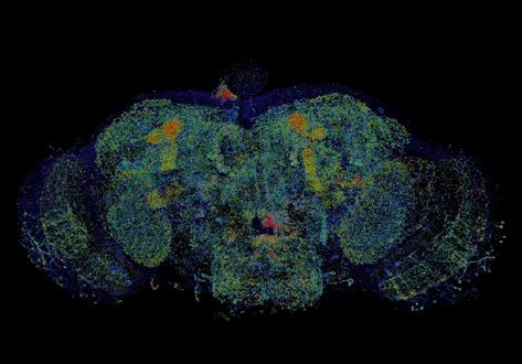
Combined expansion and lattice light sheet microscopy enables high
speed, nanoscale molecular imaging of neural circuits over large volumes.
Optical and electron microscopy have made tremendous inroads in understanding the complexity of the brain, but the former offers insufficient resolution to reveal subcellular details and the latter lacks the throughput and molecular contrast to visualize specific molecular constituents over mm-scale or larger dimensions. We combined expansio... Read more
Ruixuan Gao, Shoh M Asano, Srigokul Upadhyayula, Igor Pisarev, Daniel E Milkie, Tsung-Li Liu, Ved Singh, Austin Graves, Grace H Huynh, Yongxin Zhao, John Bogovic, Jennifer Colonell, Carolyn M Ott, Christopher Zugates, Susan Tappan, Alfredo Rodriguez, Kishore R Mosaliganti, Sean G Megason, Jennifer Lippincott-Schwartz, Adam Hantman, Gerald M Rubin, Tom Kirchhausen, Stephan Saalfeld, Yoshinori Aso, Edward S Boyden, Eric Betzig
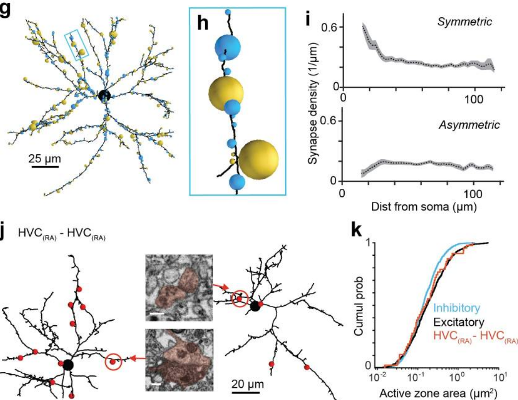
EM connectomics reveals axonal target variation in a sequence-generating network
The sequential activation of neurons has been observed in various areas of the brain, but in no case is the underlying network structure well understood. Here we examined the circuit anatomy of zebra finch HVC, a cortical region that generates sequences underlying the temporal progression of the song. We combined serial block-face electron microscopy with light microscopy to determine the cell types targeted by HVC(RA) neurons, which control song timing. Close to their soma, axons... Read more
Jörgen Kornfeld, Sam E Benezra, Rajeevan T Narayanan, Fabian Svara, Robert Egger, Marcel Oberlaender, Winfried Denk, Michael A Long